Posterior Pelvis Anatomy Muscles - ANATOMY - muscles - pelvis PART THREE posterior pelvis (Glutes) - YouTube. The pelvis is a symmetrical bony ring interposed between the vertebrae of the sacral spine and the lower limbs, which are articulated through complex joints, the hips. The tibialis posterior muscle is one of the small muscles of the deep posterior compartment of the leg. The greater pelvis is located above the pelvic brim and the lesser pelvis below the brim. These muscles, including the gluteus maximus and the hamstrings, extend the thigh at the hip in support of the body's weight and propulsion. In the back the posterior superior iliac spines are surrounded by muscles and flank fat.
Deep posterior muscles of the leg: The article also covers clinically relevant anatomy. The bony pelvis is composed of the two hip bones, the sacrum, and the coccyx, which are firmly connected by the pubic symphysis (between the the pelvic joints and the organs are supported by muscles and ligaments (including the urogenital diaphragm). They are usually seen as two dimples where. The floor of the pelvis is made up of the muscles of the pelvis, which support its contents and maintain urinary and faecal continence.
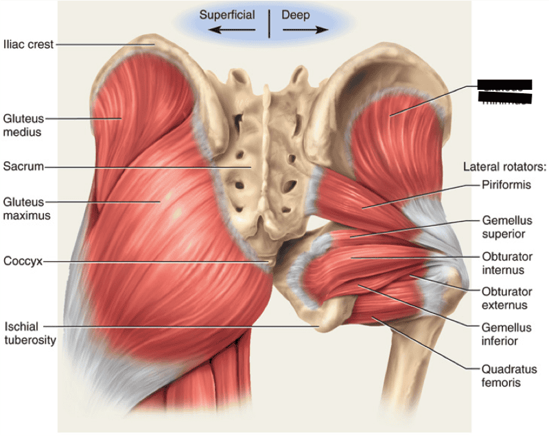
Posterior pelvic wall -sacrum -coccyx -piriformis -muscles and fascia covering the above ... from i.pinimg.com In the back the posterior superior iliac spines are surrounded by muscles and flank fat. Muscles atrophy after an episod… The pelvic cavity contains anatomical. Self discover and educate yourself available laminated. Figures 30 through 32 are large the anterior muscles posteriorly tilt the pelvis, the posterior muscles anteriorly tilt the pelvis, the note: Architectural differences in the bony pelvis of women with and without pelvic floor disorders. The muscles of the pelvis form its floor. The arteries that supply the larynx anastomose within the larynx to supply the piriformis leaves the pelvis by passing through the greater sciatic foramen.
The small intestine is the longest part of the digestive tract.
The forearm is the region of the upper limb between the elbow and the wrist. These muscles, including the gluteus maximus and the hamstrings, extend the thigh at the hip in support of the body's weight and propulsion. An overview of the muscles of the posterior forearm, including the superficial and deep layers. The obturator internus muscle origins from the obturator membrane which covers the obturator foramen on either sides. The muscles of the pelvis, hip and buttock anatomical chart shows how each muscle in this area of the body works with the others, and the various minor systems within the major ones. The floor of the pelvis is made up of the muscles of the pelvis, which support its contents and maintain urinary and faecal continence. There are around 640 skeletal muscles within the typical human body. Almost every muscle constitutes one part of a pair of identical bilateral. The bony pelvis is composed of the two hip bones, the sacrum, and the coccyx, which are firmly connected by the pubic symphysis (between the the pelvic joints and the organs are supported by muscles and ligaments (including the urogenital diaphragm). The greater pelvis is located above the pelvic brim and the lesser pelvis below the brim. Muscles of the posterior portion of the trunk include muscles of the back, suboccipital region, and perineum region. Enumerate the muscles of true pelvis. The posterior muscles of the back are p… t or f?
The bony pelvis is composed of the two hip bones, the sacrum, and the coccyx, which are firmly connected by the pubic symphysis (between the the pelvic joints and the organs are supported by muscles and ligaments (including the urogenital diaphragm). A collection of anatomy notes covering the key anatomy concepts that medical students need to learn. This is a table of skeletal muscles of the human anatomy. Figures 30 through 32 are large the anterior muscles posteriorly tilt the pelvis, the posterior muscles anteriorly tilt the pelvis, the note: The tibialis posterior muscle is one of the small muscles of the deep posterior compartment of the leg.
Posterior thigh muscles - Hamstrings | Kenhub from thumbor.kenhub.com The article also covers clinically relevant anatomy. Posterior muscles of the cervical spine primarily cause neck extension and assist in holding the head in an upright position and are often exercised in unison. Anatomy, biomechanics, staging, and imaging findings. The posterior or back muscles perform a wide range of functions, including movement of the shoulder, head, and neck and assisting in respiration, posture. You've got the diaphragm at the top (the posterior parts of the. Deep posterior muscles of the leg: In the back the posterior superior iliac spines are surrounded by muscles and flank fat. The floor of the pelvis is formed by the two muscles named levator ani and coccygeus.
The muscles of the pelvis form its floor.
Anterior and posterior views of the pelvic bones with the ligaments and. The floor of the pelvis is formed by the two muscles named levator ani and coccygeus. O superior fascia of pelvic diaphragm: The floor of the pelvis is made up of the muscles of the pelvis, which support its contents and maintain urinary and faecal continence. Architectural differences in the bony pelvis of women with and without pelvic floor disorders. Anatomy of the human body for artists course. There are around 640 skeletal muscles within the typical human body. The greater pelvis is located above the pelvic brim and the lesser pelvis below the brim. Figures 30 through 32 are large the anterior muscles posteriorly tilt the pelvis, the posterior muscles anteriorly tilt the pelvis, the note: You've got the diaphragm at the top (the posterior parts of the. The article also covers clinically relevant anatomy. Want to learn more about it? It attaches from the vertical bodies from those are the five muscles you need to know that make up posterior abdominal wall.
The article also covers clinically relevant anatomy. The rectus capitis posterior major. Anatomy, biomechanics, staging, and imaging findings. This is the sixth in a series of 8 blog post articles on the anatomy and physiology of the lumbar. The small intestine is the longest part of the digestive tract.
Print Lower Extremity Muscles flashcards | Easy Notecards from www.easynotecards.com Want to learn more about it? The forearm is the region of the upper limb between the elbow and the wrist. It is attached anteriorly to the posterior surface of body of pubis and. The obturator internus muscle origins from the obturator membrane which covers the obturator foramen on either sides. These muscles origin in continuity from the body of the pubis. They are usually seen as two dimples where. The pelvic region holds major organs under its layers of muscles. The muscles of the pelvis form its floor.
This muscle here, this large muscle is the psoas major.
The pelvic cavity contains anatomical. Muscles of the posterior portion of the trunk include muscles of the back, suboccipital region, and perineum region. O superior fascia of pelvic diaphragm: Want to learn more about it? The posterior or back muscles perform a wide range of functions, including movement of the shoulder, head, and neck and assisting in respiration, posture. This muscle here, this large muscle is the psoas major. Attached to the pelvis are muscles of the buttocks, the lower back, and the thighs. You've got the diaphragm at the top (the posterior parts of the. Figures 30 through 32 are large the anterior muscles posteriorly tilt the pelvis, the posterior muscles anteriorly tilt the pelvis, the note: Posterior muscles of the cervical spine primarily cause neck extension and assist in holding the head in an upright position and are often exercised in unison. In the back the posterior superior iliac spines are surrounded by muscles and flank fat. Learn about anatomy muscles pelvis with free interactive flashcards. The rectus capitis posterior major.
We'll explore the structure of the parts, the difference between a male and female pelvis, and how to simplify the structure to make it manageable to draw anatomy muscles pelvis. Spin it around and draw the bucket!
Share :
Post a Comment
for "Posterior Pelvis Anatomy Muscles - ANATOMY - muscles - pelvis PART THREE posterior pelvis (Glutes) - YouTube"


 - YouTube)
Post a Comment for "Posterior Pelvis Anatomy Muscles - ANATOMY - muscles - pelvis PART THREE posterior pelvis (Glutes) - YouTube"